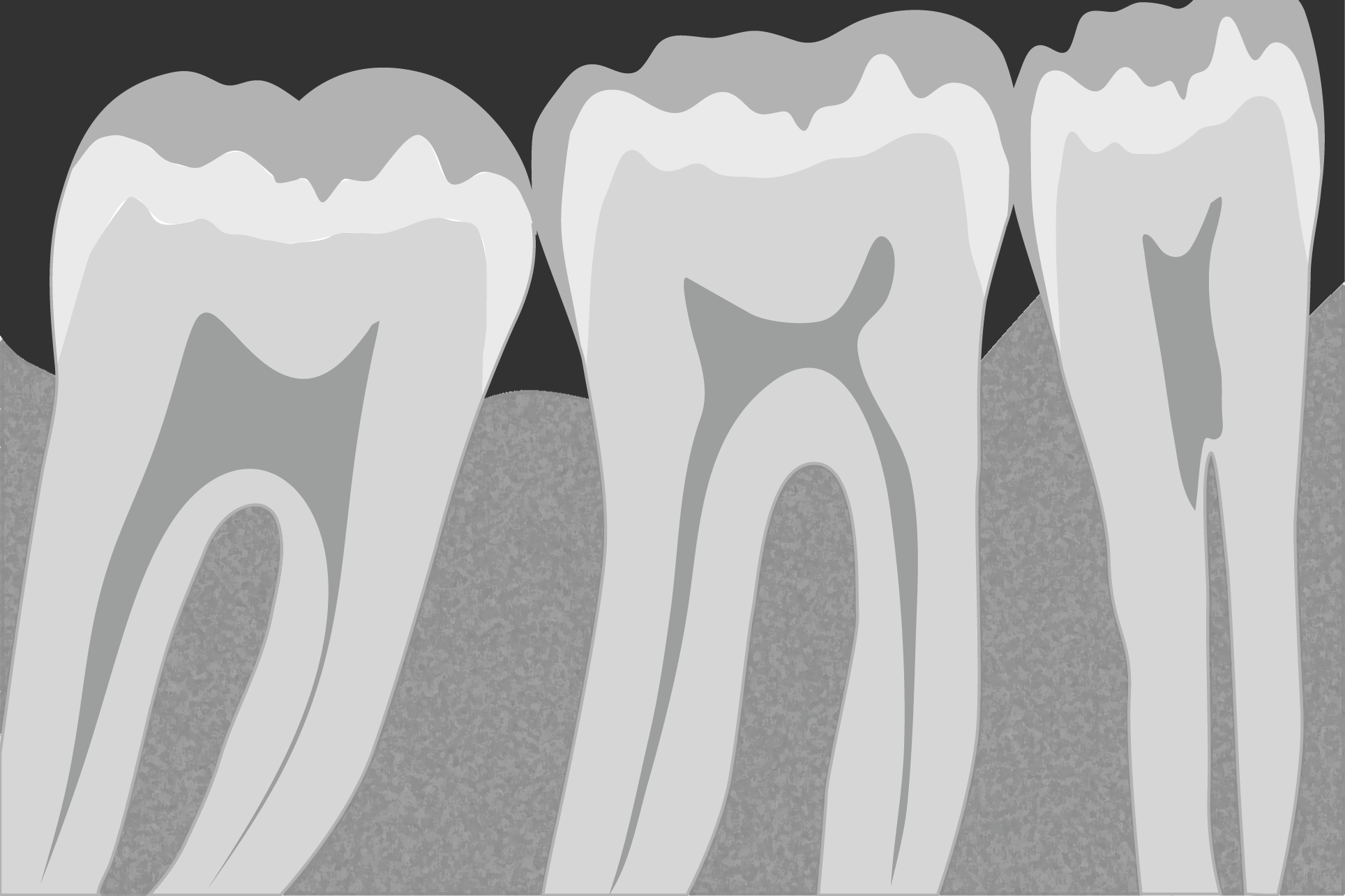
To identify an unknown implant, a clear and parallel periapical radiograph is essential. This technique provides detailed information about the implant, including the shape of the neck, the spires, the apex and in some cases allows us to distinguish the connection, providing valuable data for identification. In addition, it allows us to assess the condition of the teeth and surrounding bone, which is key in the evaluation of the treatment.
Periapical radiography is an intraoral imaging technique used in dentistry to obtain a detailed view of a specific tooth and its surrounding structure. In this technique, images of the dental crown, root and adjacent bone are captured, providing information about dental health, root anatomy and the condition of the bone around the tooth. It is performed using radiographic plates placed close to the area of interest and provides a complete view of the periapical region, which is useful for diagnosis and dental treatment planning.
What is a periapical radiograph?
TECHNIQUES FOR PERFORMING PERIAPICAL RADIOGRAPHY
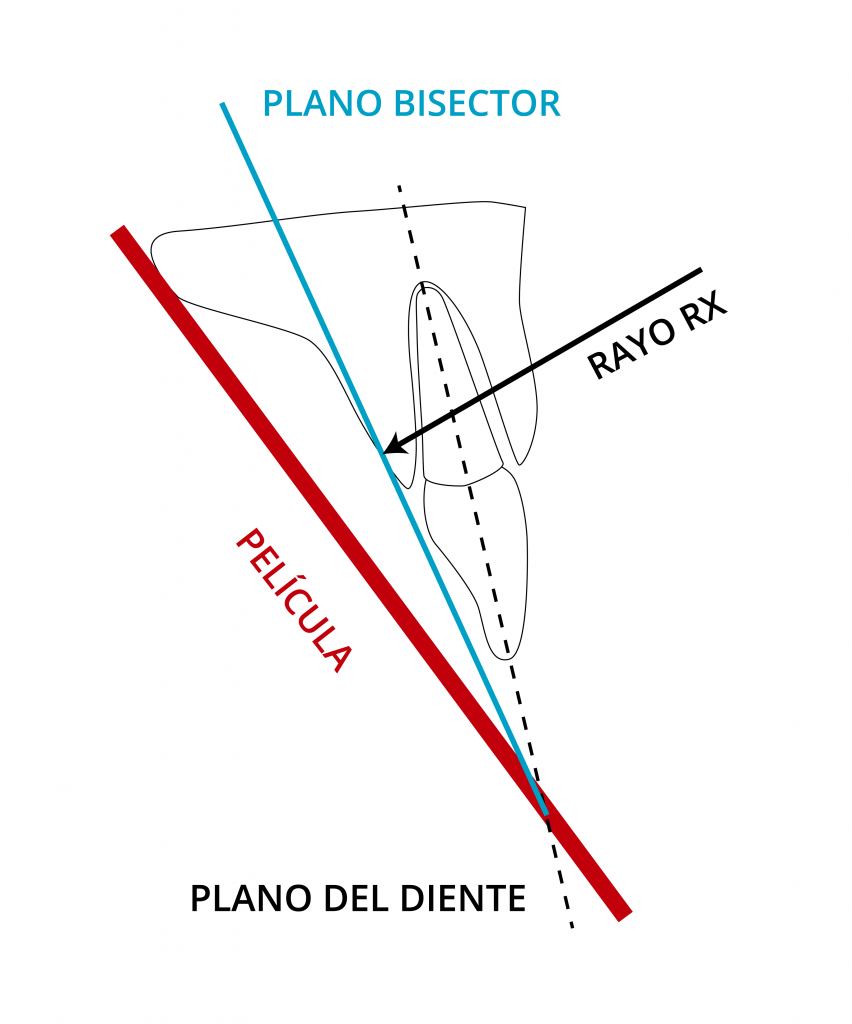
BISECTOR TECHNIQUE
The plane of the film and the axial axis of the tooth forms an angle with vertex at the point of contact of the film with the tooth. The beam is directed towards the apex of the tooth, perpendicular to the plane of the bisector.
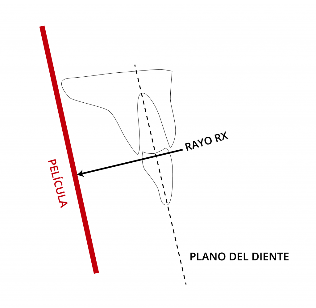
PARALLELISM TECHNIQUE
The radiographic film is kept parallel to the axial axis of the teeth. The central beam of the beam is directed perpendicularly at right angles to the teeth and the film.

PROCEDURE
1.
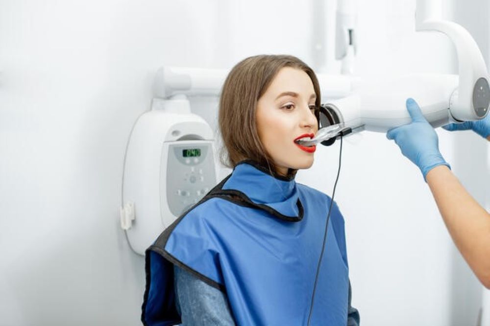
PATIENT PREPARATION
The patient is placed in the dental chair, protected by a lead apron and a thyroid collar to minimise radiation exposure.
2.
POSITIONING OF THE FILM
A special device with a rod and ring is used to hold the film in the patient’s mouth. The holder and receiver are placed in the mouth, making sure that the tooth or teeth to be studied touch the bite block, and verifying that the plane of occlusion is horizontal.
3.
ADJUSTMENT OF THE MACHINE
The locator ring is lowered until it is in contact with the patient’s face, ensuring the correct distance between the focal point and the plate. During this process, the patient is asked to maintain the proper position to capture the optimal image.
4.
X-RAY EXPOSURE
Once everything is ready, the operator activates the machine to expose the film to radiation for a brief moment, thus capturing the radiographic image required for dental diagnosis.